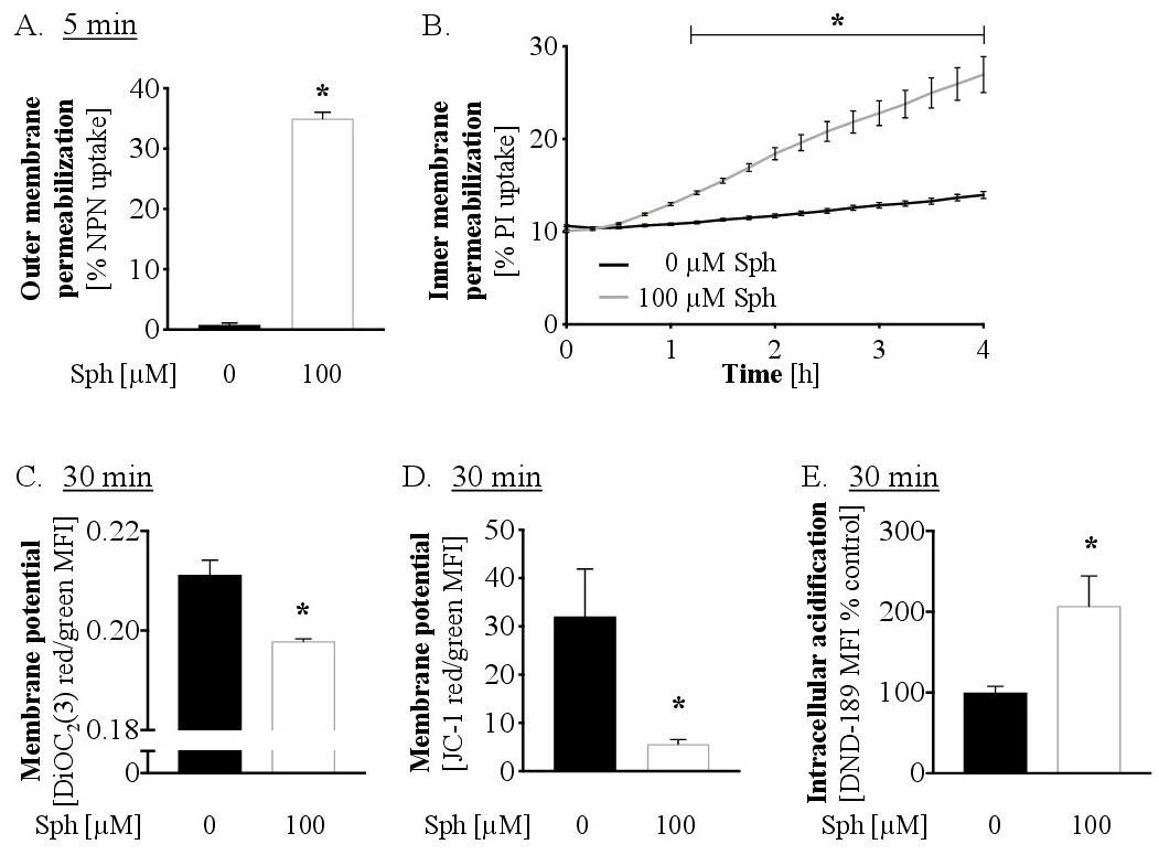
Fig. 6. Sphingosine permeabilizes the outer membrane and disrupts inner membrane potential. (A) Outer membrane permeabilization was determined by NPN uptake in P. aeruginosa 762. Bacteria were treated with normal saline or 100 µM sphingosine and NPN fluorescence measured using a microplate reader. 100 % NPN uptake was defined as the signal obtained upon treatment of bacteria with 67 % ethanol. n = 12 samples/group, spread across three independent experiments. * p<0.05 vs 0 µM (Student t test). (B) Inner membrane permeabilization was determined by PI uptake in P. aeruginosa 762. PI fluorescence was measured over 4 h after sphingosine addition using a microplate reader. 100 % PI uptake was defined as the signal obtained upon treatment of bacteria with 67 % ethanol. n = 12 samples/group, spread across three independent experiments. * p<0.05 vs 0 µM (two-way repeated-measures ANOVA with Dunnett posttest). (C+D) Membrane potential was determined by DIOC2(3). (C) and JC-1 (D) fluorescence shift from green to red using flow cytometry. Samples were analyzed 30 min after sphingosine addition. n = 6 samples/group, spread across two independent experiments. * p<0.05 vs 0 µM (Student t test). (E) Changes in intracellular pH were measured with DND-189 in P. aeruginosa 762. DND-189 fluorescence was measured by flow cytometry 30 min after sphingosine addition. n = 9 samples/group, spread across three independent experiments. * p < 0.05 vs 0 µM (Student t test).